
The Effect of Head Position on Buccal Cortical Bone Thickness Measurements in CBCT Scans: A Human Dry Mandible Study
 Abstract views: 197
/
Abstract views: 197
/  PDF downloads: 245
DOI:
https://doi.org/10.58600/eurjther1974Keywords:
CBCT, Head position, Buccal cortical bone thickness, ImplantAbstract
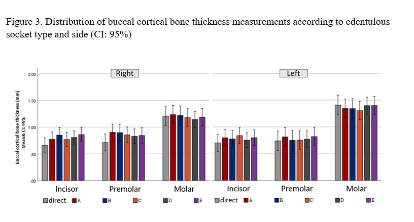
Objective: The aim of this study was to compare buccal cortical bone thickness measurements on cone beam computed tomography (CBCT) scans of human dry mandibles with direct measurements and to evaluate the effect of different head positioning on measurements.
Methods: In total, direct linear measurements were made at reference points on the buccal bone surfaces in toothless sockets in 26 human dry mandibles. CBCT scans were performed in the central position and with four different types of head position (to the right-left, to the anterior-posterior). Thickness measurements were performed on cross-sectional sections from relevant areas where heated gutta-percha was placed. Measurements were summarized as mean±standard deviation. Differences between measurements were analyzed by ANOVA and Friedmann test.
Results: Compared to direct measurements, buccal cortical bone thickness in CBCT scans was higher in the incisor and premolar regions, while lower values were obtained in the molar region. These differences were statistically significant but less than 0.2 mm (p<0.005). Different head positions had no effect on measurements on CBCT images (p>0.005). Intraobserver agreement for buccal bone thickness was found to be high (ICC=0.902-0.976).
Conclusion: It demonstrated a clinically acceptable difference between direct measurements and CBCT measurements of mandibular buccal cortical bone thickness. Additionally, no differences in measurements were observed between different types of head positions.
Metrics
References
Scarfe WC, Farman AG, Sukovic P (2006) Clinical applications of cone beam computed tomography in dental practice. J Can Dent Assoc. 72(1):75-80.
Kim MK, Kang SH, Lee EH, Lee SH, Park W (2012) Accuracy and validity of stitching sectional cone beam computed tomographic images. J Craniofac Surg. 23(4):1071-1076. https://doi.org/10.1097/SCS.0b013e31824e2c85
Torres MG, Campos PS, Segundo NP, Navarro M, Crusoé Rebello I (2012) Accuracy of linear measurements in cone beam computed tomography with different voxel sizes. Implant Dent. 21 (2):150-155. https://doi.org/10.1097/ID.0b013e31824bf93c
Cavalcanti MG, Rocha SS, Vannier MW (2004) Craniofacial measurements based on 3D-CT volume rendering: implications for clinical applications. Dentomaxillofac Radiol. 33(3):170-176. https://doi.org/10.1259/dmfr/13603271
Wood R, Sun Z, Chaudhry J, Tee BC, Kim DG, Leblebicioglu B, England G (2013) Factors affecting the accuracy of buccal alveolar bone height measurements from cone-beam computed tomography images. Am J Orthod Dentofacial Orthop. 143(3):353-363. https://doi.org/10.1016/j.ajodo.2012.10.019
Dantas LL, Ferreira PP, Oliveira L, Neves FS, Campos PSF, Scarfe WC, I Crusoe-Rebello (2019) Cone beam computed tomography devices in the evaluation of buccal bone in anterior teeth. Aust Dent J. 2019;64(2):161-166. https://doi.org/10.1111/adj.12685
Van Dessel J, Nicolielo LF, Huang Y, Coudyzer W, Salmon B, Lambrichts I, Jacobs R (2017) Accuracy and reliability of different cone beam computed tomography (CBCT) devices for structural analysis of alveolar bone in comparison with multislice CT and micro-CT. Eur J Oral Implantol. 10(1):95-105.
Dings JPJ, Verhamme L, Merkx MA, Xi T, Meijer GJ, Maal TJ (2019) Reliability and accuracy of cone beam computed tomography versus conventional multidetector computed tomography for image-guided craniofacial implant planning: an in vitro study. Int J Oral Maxillofac Implants. 34(3):665-672. https://doi.org/10.11607/jomi.6915
Salimov F, Tatli U, Kürkçü M, Akoglan M, Oztunç H, Kurtoglu C (2014) Evaluation of relationship between preoperative bone density values derived from cone beam computed tomography and implant stability parameters: a clinical study. Clin Oral Implants Res. 25(9):1016-1021. https://doi.org/10.1111/clr.12219
Stamatakis HC, Steegman R, Dusseldorp J, Ren Y (2019) Head positioning in a cone beam computed tomography unit and the effect on accuracy of the three-dimensional surface mode. Eur J Oral Sci. 127(1):72-80. https://doi.org/10.1111/eos.12582
Ludlow JB, Laster WS, See M, Bailey LJ, Hershey HG (2007) Accuracy of measurements of mandibular anatomy in cone beam computed tomography images. Oral Surg Oral Med Oral Pathol Oral Radiol Endod. 103(4):534-542. https://doi.org/10.1016/j.tripleo.2006.04.008
Hassan B, van der Stelt P, Sanderink G (2009) Accuracy of three-dimensional measurements obtained from cone beam computed tomography surface-rendered images for cephalometric analysis: influence of patient scanning position. Eur J Orthod. 31(2):129-134. https://doi.org/10.1093/ejo/cjn088
El-Beialy AR, Fayed MS, El-Bialy AM, Mostafa YA (2011) Accuracy and reliability of cone-beam computed tomography measurements: Influence of head orientation. Am J Orthod Dentofacial Orthop. 140(2):157-165. https://doi.org/10.1016/j.ajodo.2010.03.030
Shokri A, Miresmaeili A, Farhadian N, Falah-Kooshki S, Amini P, Mollaie N (2016) Effect of head position on maxillofacial transverse measurements made on the skull and cone beam computed tomography scans. Braz Dent J. 27(5):604-608. https://doi.org/10.1590/0103-6440201601166
Kim JH, Jeong HG, Hwang JJ, Lee JH, Han SS (2016) The impact of reorienting cone-beam computed tomographic images in varied head positions on the coordinates of anatomical landmarks. Imaging Sci Dent. 46(2):133-139. https://doi.org/10.5624/isd.2016.46.2.133
Nascimento MDCC, Boscolo SMA, Haiter-Neto F, Santos ECD, Lambrichts I, Pauwels R, Jacobs R (2017) Influence of basis images and skull position on evaluation of cortical bone thickness in cone beam computed tomography. Oral Surg Oral Med Oral Pathol Oral Radiol. 123(6):707-713. https://doi.org/10.1016/j.oooo.2017.01.015
Caldas MdeP, Ramos-Perez FM, de Almeida SM, Haiter-Neto F (2010) Comparative evaluation among different materials to replace soft tissue in oral radiology studies. J Appl Oral Sci. 18(3):264-267. https://doi.org/10.1590/s1678-77572010000300012
Li L, Wu D, Liu P, Liang L, Chen Z (2011) Experimental measurement of human head motion for clinical dental CBCT system design. IEEE Nuclear Science Symposium Conference Record, 2895-2897. https://doi.org/10.1109/NSSMIC.2011.6152513
Lee KM, Song JM, Cho JH, Hwang HS (2016) Influence of Head Motion on the Accuracy of 3D Reconstruction with Cone-Beam CT: Landmark Identification Errors in Maxillofacial Surface Model. PLoS ONE 11(4):e0153210. https://doi.org/10.1371/journal.pone.0153210
Al-Haj Husain A, Stadlinger B, Özcan M, Schönegg D, Winklhofer S, Al-Haj Husain N, Piccirelli M, Valdec S (2023) Buccal bone thickness assessment for immediate anterior dental implant planning: A pilot study comparing cone-beam computed tomography and 3D double-cho steady-state MRI. Clin Implant Dent Relat Res. 25(1):35-45. https://doi.org/10.1111/cid.13160
Cetmili H, Tassoker M, Sener S (2019) Comparison of cone-beam computed tomography with bitewing radiography for detection of periodontal bone loss and assessment of effects of different voxel resolutions: an in vitro study. Oral Radiol. 35(2):177-183. https://doi.org/10.1007/s11282-018-0336-x
Alkan BA, Aral CA, Aral K, Acer N, Şişman Y (2016) Quantification of circumferential bone level and extraction socket dimensions using different imaging and estimation methods: a comparative study. Oral Radiol. 32(3):145-153. https://doi.org/10.1007/s11282-015-0225-5
Timock AM, Cook V, McDonald T, Leo MC, Crowe J, Benninger BL, Covell DA Jr (2011) Accuracy and reliability of buccal bone height and thickness measurements from cone-beam computed tomography imaging. Am J Orthod Dentofacial Orthop. 140(5):734-744. https://doi.org/10.1016/j.ajodo.2011.06.021
Damstra J, Fourie Z, Huddleston Slater JJ, Ren Y (2010) Accuracy of linear measurements from cone-beam computed tomography-derived surface models of different voxel sizes. Am J Orthod Dentofacial Orthop. 137(1):16.e1-17. https://doi.org/10.1016/j.ajodo.2009.06.016
Al Abbady NA, Hamdy RM, El Dessouky SH (2019) Accuracy of linear measurements using low dose cone beam computed tomography protocol versus direct skull linear measurements: An in vitro study [version 1; peer review: 2 not approved]. F1000Research 8:25 https://doi.org/10.12688/f1000research.17607.1
Kamburoğlu K, Kolsuz E, Kurt H, Kiliç C, Özen T, Paksoy CS (2011) Accuracy of CBCT measurements of a human skull. J Digit Imaging. 24(5):787-793. https://doi.org/10.1007/s10278-010-9339-9
Ozemre MO, Gulsahi A (2018) Comparison of the accuracy of full head cone beam CT images obtained using a large field of view and stitched images. Dentomaxillofac Radiol. 47(7):20170454. https://doi.org/10.1259/dmfr.20170454
Nikneshan S, Aval SH, Bakhshalian N, Shahab S, Mohammadpour M, Sarikhani S (2014) Accuracy of linear measurement using cone-beam computed tomography at different reconstruction angles. Imaging Sci Dent. 44(4):257-262. https://doi.org/10.5624/isd.2014.44.4.257
Leung CC, Palomo L, Griffith R, Hans MG (2010) Accuracy and reliability of cone-beam computed tomography for measuring alveolar bone height and detecting bony dehiscences and fenestrations. Am J Orthod Dentofacial Orthop. 137(4):109-119. https://doi.org/10.1016/j.ajodo.2009.07.013
Lund H, Gröndahl K, Gröndahl HG (2009) Accuracy and precision of linear measurements in cone beam computed tomography Accuitomo tomograms obtained with different reconstruction techniques. Dentomaxillofac Radiol. 38(6):379-386. https://doi.org/10.1259/dmfr/15022357
31.Kamburoğlu K, Ereş G, Akgün C, Yeta EN, Gülen O, Karacaoĝlu F (2015) Effect of voxel size on accuracy of cone beam computed tomography- aided assessment of periodontal furcation involvement. Oral Surg Oral Med Oral Pathol Oral Radiol. 120(5):644-650. https://doi.org/10.1016/j.oooo.2015.07.030
Kolsuz ME, Bagis N, Orhan K, Avsever H, Demiralp KÖ (2015) Comparison of the influence of FOV sizes and different voxel resolutions for the assessment of periodontal defects. Dentomaxillofac Radiol. 44(7):20150070. https://doi.org/10.1259/dmfr.20150070
Downloads
Published
How to Cite
License
Copyright (c) 2024 European Journal of Therapeutics

This work is licensed under a Creative Commons Attribution-NonCommercial 4.0 International License.
The content of this journal is licensed under a Creative Commons Attribution-NonCommercial 4.0 International License.


