
Imaging of the Ethmomaxillary Sinus, its Prevalence, and Evaluation of its Relationship with Chronic Rhinosinusitis
 Abstract views: 258
/
Abstract views: 258
/  PDF downloads: 198
DOI:
https://doi.org/10.58600/eurjther1891Keywords:
Ethmoid Sinus, Maxillary Sinus, Paranasal Sinuses, Sinusitis, TomographyAbstract
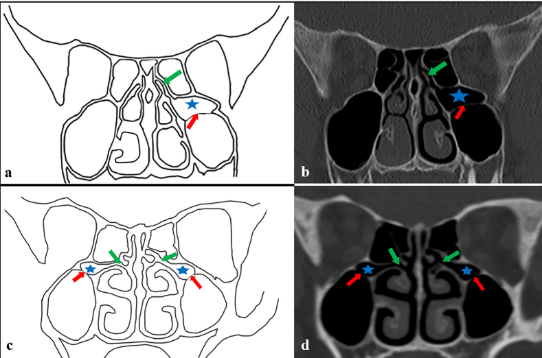
Objective: The presence of an ethmomaxillary sinus (EMS) may increase the susceptibility to inflammatory paranasal sinus diseases such as chronic rhinosinusitis (CRS) and cause difficulties in surgical interventions to the paranasal sinuses. Therefore, this study aimed to examine the EMS in patients with and without CRS.
Methods: The study included 150 patients (300 sides) diagnosed with CRS by the ear–nose–throat clinic and 151 individuals (302 sides) without CRS. Paranasal sinus computed tomography images were reviewed retrospectively. The presence of an EMS (bilateral or not) and its relationship with age and sex were examined. The severity of CRS was determined with the Lund–Mackay scoring system, and its relationship with EMS was evaluated.
Results: The EMS was detected in 7 patients (7/301, 2.32%) and 9 sides (9/602, 1.49%) of 301 patients (602 sides) included. The incidence in the CRS group was 2.6%. Three cases were unilateral, and one was bilateral. The incidence in the control group was 1.98%, two cases were unilateral, and one was bilateral. According to the Lund–Mackay scoring system, the mean CRS severity was 8.62 (±5.47). Its severity was 5.25 (±3.94) in the EMS group and 8.71 (±5.48) in the non-EMS group.
Conclusion: No statistically significant difference was found between the groups with and without CRS in terms of the presence of EMS (p = 0.723). No evidence reveals that EMS increased the severity of CRS.
Metrics
References
Snell RS (2011) Clinical anatomy by regions, 9th edn. Lippincott Williams & Wilkins, Philadelphia
Standring S (2021) Gray's anatomy e-book: the anatomical basis of clinical practice, 42nd edn. Elsevier Health Sciences, London
Kamdi P, Nimma V, Ramchandani A, Ramaswami E, Gogri A, Umarji H (2018) Evaluation of haller cell on CBCT and its association with maxillary sinus pathologies. J Indian Acad Oral Med Radiol. 30:41-45. https://doi.org/10.4103/jiaomr.jiaomr_22_18
Chmielik LP, Chmielik A (2017) The prevalence of the Onodi cell – Most suitable method of CT evaluation in its detection. Int J Pediatr Otorhinolaryngol. 97:202-205. https://doi.org/https://doi.org/10.1016/j.ijporl.2017.04.001
Cao C, Zhou F, Song Z, Tao Z, Xu Y (2020) Computed Tomography Image Analysis and Clinical Correlations of Retromaxillary Cells. Ear Nose Throat J. 2022;101(7):435-442. https://doi.org/10.1177/0145561320936963
Behboudi H, Heirati A, Behboudi H, Akbari M, Ramezani N, Shalchizadeh M, Hajian A, Nemati S (2021) Nasolacrimal Duct Obstruction and Frequency of Agger Nasi Cell and other Anatomical Field Variations: A Controlled Study in Northern Iran. Acta Med Iran. 59(4):203. https://doi.org/10.18502/acta.v59i4.6218
Zhou F, Cao C, Fan W, Tan L, Liu P, Lv H, Xu Y (2021) The imaging anatomy of ethmomaxillary sinus and its impact on chronic rhinosinusitis. Eur Arch Otorhinolaryngol. 278(3):719-26. https://doi.org/10.1007/s00405-020-06322-y
Jinfeng L, Jinsheng D, Xiaohui W, Yanjun W, Ningyu W (2017) The Pneumatization and Adjacent Structure of the Posterior Superior Maxillary Sinus and Its Effect on Nasal Cavity Morphology. Med Sci Monit 23:4166-4174. https://doi.org/10.12659/msm.903173
Liu J, Dai J, Wen X, Wang Y, Zhang Y, Wang N (2018) Imaging and anatomical features of ethmomaxillary sinus and its differentiation from surrounding air cells. Surg Radiol Anat. 40(2):207-15. https://doi.org/10.1007/s00276-018-1974-8
Şirikçi A, Bayazıt YA, Bayram M, Kanlıkama M (2004) Ethmomaxillary sinus: a particular anatomic variation of the paranasal sinuses. Eur Radiol. 14:281-285. https://doi.org/10.1007/s00330-003-1993-6
Poojary N, Meghanandh KR, Patil T (2022) Posterior Ethmomaxillary Cells: Anatomical Variation to be Considered in Endoscopic Sinus Surgery. Indian J Otolaryngol Head Neck Surg. 1-4. https://doi.org/10.1007/s12070-022-03298-5
Gore MR, Ebert CS, Jr., Zanation AM, Senior BA (2013) Beyond the "central sinus": radiographic findings in patients undergoing revision functional endoscopic sinus surgery. Int Forum Allergy Rhinol 3:139-146. https://doi.org/10.1002/alr.21079
Chen JJ, Chen D-L, Chen C-J (2011) The Lund-Mackay score for adult head and neck computed tomography. J Radiol Sci 36:203-208.
Herzallah IR, Saati FA, Marglani OA, Simsim RF (2016) Retromaxillary Pneumatization of Posterior Ethmoid Air Cells: Novel Description and Surgical Implications. Otolaryngol Head Neck Surg 155:340-346. https://doi.org/10.1177/0194599816639943
Ozcan KM, Selcuk A, Oruk V, Sarikaya Y, Dere H (2008) Ethmomaxillary sinus. Eur Arch Otorhinolaryngol. 265:185–188. https://doi.org/10.1007/s00405-007-0444-4
Melnichenko YM, Savrasova N, Kabak S, Mekhtiev R (2022) Anatomical variations of the ethmomaxillary sinus. Vestn Otorinolaringol. 87(3):46-50. http://doi.org/10.17116/otorino20228703146
Kim SJ, Moon JW, Lee HM (2023) Clinical and imaging features of ethmomaxillary sinus compared to Haller’s cell. Eur Arch Otorhinolaryngol. 280(12): 5401-5406. https://doi.org/10.1007/s00405-023-08148-w
Downloads
Published
How to Cite
License
Copyright (c) 2023 European Journal of Therapeutics

This work is licensed under a Creative Commons Attribution-NonCommercial 4.0 International License.
The content of this journal is licensed under a Creative Commons Attribution-NonCommercial 4.0 International License.


