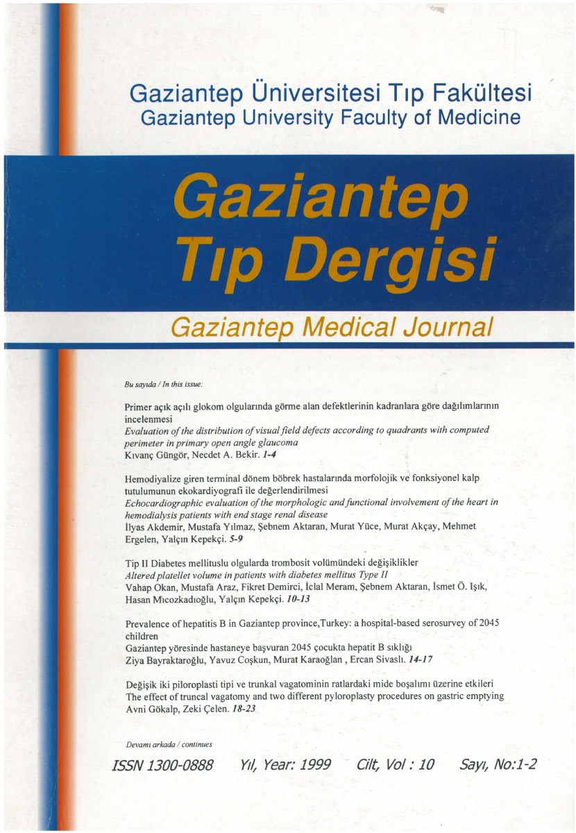
Evaluation of the distribution of visual field defects according to quadrants by computed perimetry in primary open angle glaucoma
DOI:
https://doi.org/10.58600/eurjther.1999-10-1-2-1468-archKeywords:
Primary open angle glaucoma, computed perimetry, visual field defectsAbstract
in this study, our aim was to research early term changes of visual field in primary open angle glaucoma (POAG) if there are any tendencies to localize for superior, inferior - half and quadrants of superior-temporal, superior-nasal, inferior-temporal and inferior-nasal.
We evaluated right eyes of 41 cases with primary open angle glaucoma which were 19 female of those and 22 male of those. Routine ophthalmic examinalion and automated perimetry was performed and determined with the G 1 program in the Octopus 500 EZ perimeter by the same person. The mean and standard deviations of'averaged mean defects"for superior, inferior - halt and quadrants of superior-temporal, nasal or inferior, temporal-nasal were compared in POAG cases. When we compared "averaged mean defects", the difference was statistically significant in visual fields of superior-half and superior-temporal quadrant (p<0.05).
We concluded that visual field defects in primary open angle glaucoma cases were localized in superior-half which probably shows the role of vascular factors in the ethiopathogenesis of glaucoma and determination of the tendencies for quadrants in visual fields need longer term studies.
Metrics
References
Shields MB. Textbook of glaucoma. (3rd ed). Baltimore, William & Wilkins, 1994; l: 1.
Flammer J, Jenni A, Bebie H, Keller B. Octopus Gi program. Glaucoma 1986; 9:67-72.
Turaçlı ME. Primer açık açılı glokomda bilgisayarlı görme alanı. XXVIII. Ulusal Türk Oftalmoloji Bülteni, Antalya, 1994; Cilt 1: 117-122.
Yedigöz N, Karatum F, Sürel Z, Aras C, Üstündağ C, Konya HE. Başlangıç glokom olgularında otomatik ve kompüterize perimetrinin yeri. Türk Oftalmoloji Gazetesi_1990; 20:475-479.
Michelson G, Langhans MJ, Groh MJM: Perfusion of the juxtapapillary retina and the neuroretinal rim area in primary open angle glaucoma. J Glaucoma 5: 91-98, 1966.
Boehm AG, Pillunat LE, Koeller U, Katz B, Schicketanz C, Klemm M, et al. regional distribution of optic nerve head blood flow. Graefe's Arch Clin Exp Ophthalmol 1999; 237:484-488.
Zeiter HJ, Shin HD, Baek NH. Visual field defects in diabetic patients with prirnary open-angle glaucoma. Am J Ophthalmol 1991; 111:581-584.
Spaeth GL. Developrnent of Glaucornatous changes of the optic nerve. in: Varma R, Spaeth GL, and Parker KW (eds) The Optic Nerve in Glaucoma. Philadelphia, JB Lippincott Co, 1993; 63-81.
Zeiter HJ, Shin HD, Juzych MS, Jarvi TS, Spoor TC, Zwas F. Visual field defects in patients with normal-tension glaucorna and patients with hightension glaucorna. Am J Ophthalmol 1992; 114:758- 763.
Caprioli J, Spaeth GL. Comparison of visual field defects in the low-tension glaucomas with those in the high-tension glaucomas. Am J Ophthalmol 1984; 97 :730-737.
King D, Drance SM, Douglas GR, Schulzer M, Wijsman K. Comparison of visual field defects in normal-tension glaucoma and high-tension glaucoma. Am J Ophthalmol 1986; 101:204-207.
Lewis RA, Hayreh SS, Phelps CD. Optic disc and visual field correlations in primary open-angle glaucoma and low-tension glaucoma. Am J Ophthalmol 1983; 96:148-152
Mikelberg F, Drance SM. Glaucomatous visual field defects. in: Ritch R, Shields MB, Krupin T (eds) The Glaucomas (2nd ed). St. Luis, The CV Mosby Co, 1996; 523-527.
Quigley HA, Hohmann RM, Addicks EM. Morphological changes in the lamina cribrosa correlated with neural loss in open-angle glaucoma. Am J Ophthalmol 1983; 95:673-681.
Quigley HA, Addicks EM, Green WR. Optic nerve damage in human glaucoma. III. Quantitative correlation of nerve fiber loss and visual field defect in glaucoma, ischemic neuropathy, papilledema and toxic neuropathy. Arch Ophthalmol 1982; 99:2159- 2262.
Downloads
Published
How to Cite
Issue
Section
License
Copyright (c) 2023 European Journal of Therapeutics

This work is licensed under a Creative Commons Attribution-NonCommercial 4.0 International License.
The content of this journal is licensed under a Creative Commons Attribution-NonCommercial 4.0 International License.


